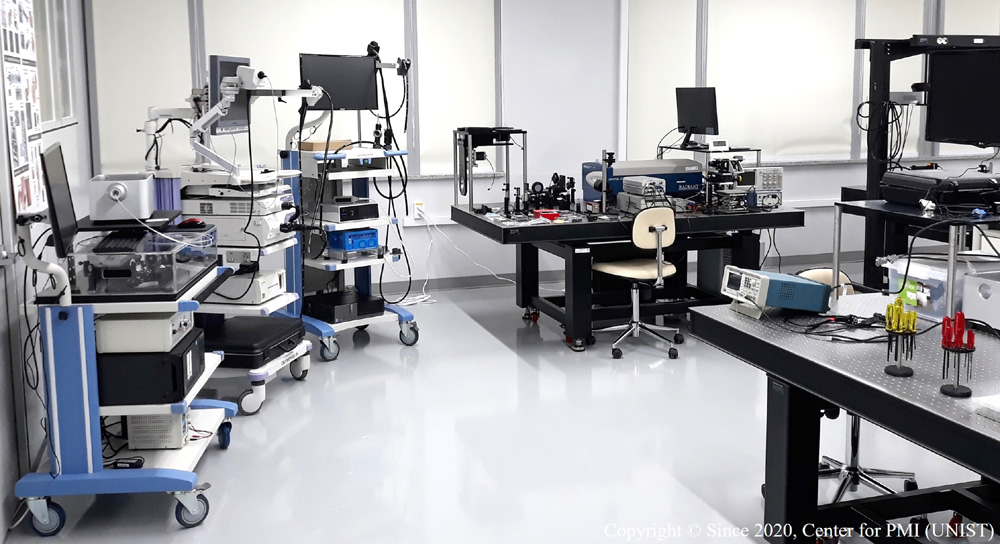
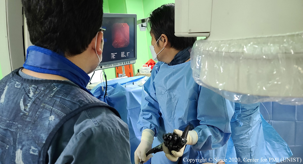
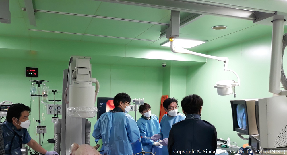
We develop various forms of novel biomedical imaging and sensing techniques based on optical and acoustical (i.e., ultrasound) methods to solve pending or unaddressed biological and medical problems. Pursuing this goal, our research needs to follow all the stages in developing the related technologies—i.e., from system design to implementation and related experiments—to demonstrate their utility and explore new application areas. Moreover, we collaborate with many specialized biology research groups in UNIST and also perform various clinical experiments with medical doctors in neighboring hospitals or medical schools throughout the country. In addition to application-oriented studies, we will also make efforts to conduct a significant amount of fundamental research to solidify the related technical basis.
Among the many existing sub-areas of optics and acoustics, we currently utilize photoacoustic and ultrasound imaging as the primary technical platforms to realize the goal. Based on the two techniques, we intend to develop new and advanced versions of imaging systems either in a separate (i.e., single-mode) or combined (i.e., dual-mode) form. In terms of combined imaging systems, our plans include a variety of miniaturized imaging devices, such as endoscopic probes for gastroenterology, intravascular imaging catheters for interventional cardiology, and many other minimally invasive imaging probes for urologic applications. We are also interested in developing handheld or portable imaging devices for dermatological treatments and a new generation of small-animal whole-body imaging and brain imaging systems that can visualize anatomical sites that cannot be covered by conventional systems.
Photoacoustic imaging, also termed photoacoustic tomography (PAT) or optoacoustic tomography (OAT), is a new technique that combines the high contrast benefit of optical imaging and the high-resolution and deep imaging capability of ultrasound imaging. In recent years, PAT has been widely recognized as one of the most promising approaches in clinical applications, such as human brain or breast imaging, minimally invasive imaging (i.e., endoscopy), and basic research, in fields such as neuroscience and cancer biology. As the technique enables high-resolution tomographic imaging at a clinically relevant depth and provides unique molecular, physiological, and functional information, it is expected that it will mature into a mainstream clinical imaging modality alongside magnetic resonance imaging (MRI), X-ray computed tomography (CT), positron emission tomography (PET), and ultrasound imaging. Furthermore, PAT is likely to become an indispensable complement to conventional optical microscopy tools, such as confocal microscopy, two-photon microscopy, and optical coherence tomography.


